
Technical Guidance Document — April 13, 2026
When referencing this document, please use the following format:
SLN Biomarker Committee. Small Lung Nodule Assessment in CT Screening Profile – 2026. Technical Guidance Document, April 13, 2026. Quantitative Medical Imaging Coalition (QMIC).
Co-chairs: Sean Fain, Artit Jirapatnakul, James Mulshine
Small Lung Nodule Biomarker Committee Members: Ricardo Avila, Raul Estepar, Ella Kazerooni, Mathis Konrad, Nancy Obuchowski, Anthony Reeves, Jessica Sieren, Mario Silva, David Yankelevitz, and Gudrun Zahlmann
Sponsored by: The American Lung Association
Member of: The Quantitative Medical Imaging Coalition and Technical Biomarker Committee
In collaboration with: Strengthening the Screening of Lung Cancer in Europe (SOLACE)
This document is a continuation of the RSNA/QIBA CT Small Lung Nodule Profile that has been improved and refined by the Small Lung Nodule Profile guideline group (slnprofile.org). The American Lung Association and QMIC supports and endorses this initiative.
1. Executive Summary
This document outlines guidance recommended by the SLN Profile for Small Lung Nodule Volume Assessment and Monitoring in Low Dose CT Screening. This Profile focuses on improving the consistency and reliability of quantitative CT volume measurements for solid lung nodules, aiding in the management of screen-detected nodules and optimizing patient care. This Profile provides a framework to ensure reliable and consistent CT imaging results across different imaging platforms and clinical sites.
The SLN Profile aims to achieve a useful level of performance for a specific biomarker. It consists of:
- The Claim (Section 2): Describes the biomarker performance.
- Conformance Checklist (Section 3): Outlines activities that generate the biomarker and sets requirements for Actors (Radiographers/technologists).
This Profile focuses on the accuracy and precision of quantitative CT volumetry for solid lung nodules between 6–10 mm in diameter. It sets requirements for:
- Acquisition Devices
- Technologists/Radiographers
- Radiologists
- Image Analysis Tools
These requirements cover various activities, from equipment quality assurance to image analysis, aiming to achieve accurate measurements and reduce unnecessary variability. The Profile provides two sets of claims:
- 95% confidence intervals for volumetric measurements of 6–10 mm solid lung nodules.
- Guidance on:
- Percentage of volumetric change needed to confirm true solid lung nodule growth with 95% confidence.
- 95% confidence interval for volumetric size change measurements.
This document is a resource for clinicians, imaging staff, vendors, purchasers, and researchers involved with pulmonary nodule volume measurements. This Profile document includes a conformance test that can be performed to test the fundamental imaging performance characteristics of the CT scanner to be used at a clinical site.
Note: This Profile sets requirements to achieve the claim, not standards of care. Patient care always takes precedence over meeting Profile goals.
2. Clinical Context and Claims
Clinical Context
This Profile focuses on quantifying volumes and volume changes of solid lung nodules with a longest bi-dimensional diameter between 6 mm and 10 mm.
Claims
When all relevant staff and equipment conform to this Profile, the following claims are supported:
Claim 1: Nodule Volume
For a measured nodule volume Y, the 95% confidence interval for the true nodule volume is Y ± (1.96 × Y × CV), where CV is the Coefficient of Variation from Table 1.
Claim 2: Nodule Volume Change
a) A measured nodule volume percentage change X indicates a true change if X > (2.77 × CV1 × 100), with 95% confidence.
b) For volume measurements Y1 and Y2 at two time points, with corresponding CVs (CV1 and CV2) from Table 1, the 95% confidence interval for volume change Z = (Y2 − Y1) ± 1.96 × √([Y1 × CV1]2 + [Y2 × CV2]2).
Claim Conditions
These claims are valid when:
- The nodule is completely solid
- Nodule longest dimension is 6–10 mm at the first time point
- Nodule’s shortest diameter is at least 60% of its longest diameter
- Nodule is measurable at both time points
- Interpolation is used for CV values between provided table values
Table 1: Coefficients of Variation (CV)
| Nodule Diameter (mm) | Nodule Volume (mm3) | CV | 95% CI Limits (mm3) | Minimum Detectable Difference |
|---|---|---|---|---|
| 6 | 113 | 0.290 | ± 64 | 80.4% |
| 7 | 154 | 0.227 | ± 68 | 62.8% |
| 8 | 268 | 0.186 | ± 98 | 51.6% |
| 9 | 382 | 0.159 | ± 119 | 43.9% |
| 10 | 524 | 0.138 | ± 142 | 38.3% |
| 11 | 697 | 0.123 | ± 168 | 34.0% |
| 12 | 905 | 0.111 | ± 196 | 30.6% |
| 13 | 1150 | 0.101 | ± 227 | 27.9% |
| 14 | 1437 | 0.093 | ± 261 | 25.6% |
| 15 | 1767 | 0.086 | ± 296 | 23.7% |
For a more detailed explanation of the clinical context, including the benefits and challenges of low-dose CT for lung nodule measurement, please refer to the full discussion in Appendix A.
For detailed examples and clinical interpretations of Claims 1 and 2, please refer to Appendix B. A web-based calculator for computing the equations in the Claims is available at https://services.accumetra.com/NoduleCalculator.html.
3. Clinical Site Conformance Checklist
This checklist provides a streamlined approach for clinical sites to achieve conformance with this Profile. It is designed to be applicable across different regions, including Europe and the USA. The checklist is divided into two main sections: Preparation and Performance. Each step indicates the responsible actor(s) in parentheses.
For detailed technical information on Profile requirements, please refer to Section 4.
Preparing for Lung Nodule Measurement
1. CT Scanner and Lung Nodule Analysis Software Verification (Radiologist)
- ☐ 1.1 Verify that the CT scanner model is approved for use in your region.
- ☐ 1.2 Verify that the volumetric software name and version are approved for use in your region.
2. CT QA and Lung Screening Protocol Verification (Medical Physicist, Technologist/Radiographer)
- ☐ 2.1 Establish and save a CT lung cancer screening protocol
- ☐ 2.2 Scan a reference object using the saved protocol
- ☐ 2.3 Obtain a passing automated CT image quality report
Performing Lung Nodule Measurement
4. CT Data Acquisition, Lung Nodule, and Segmentation Verification (Radiologist)
- ☐ 4.1 Verify same CT scanner and protocol used for follow-up scans
- ☐ 4.2 Confirm no IV contrast was used
- ☐ 4.3 Verify nodule characteristics (solid, 6–10 mm longest diameter at baseline, ≤ 1/3 attached)
- ☐ 4.4 Check for absence of significant artifacts and excessive noise
- ☐ 4.5 Verify absence of segmentation errors
Note: You can use the approved nodule calculator for measurement error guidance.
Note: Specific regional requirements (e.g., CTDIvol limits) should be incorporated into the relevant steps as needed.
4. Profile Activities
This Profile outlines the activities necessary for proper lung cancer screening to achieve the applicability of the profile claims. It is organized by key actors involved in the screening process. Each actor is responsible for specific activities that contribute to the overall quality and consistency of the screening process. Figure 1 provides a quick overview of how the different activities for CT tumor volumetry are sequenced. The following table (Table 2) outlines the actors and their required activities, with references to detailed specifications in subsequent sections.
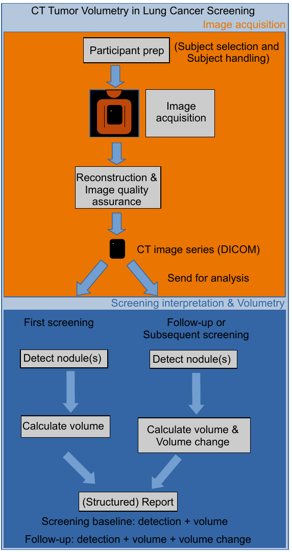
Figure 1: CT Tumor Volumetry – Activity Sequence
Table 2: Actors and Required Activities
| Actor | Activities | Section |
|---|---|---|
| Acquisition Device | Product Validation | 4.1 |
| Image Analysis Tool | Product Validation | 4.1 |
| Radiographer | Staff Qualification, Protocol Design, Subject Handling, Image Data Acquisition, Image Data Reconstruction, Image Quality Assurance | 4.2, 4.4, 4.6, 4.7, 4.8, 4.9 |
| Radiologist | Staff Qualification, Protocol Design, Subject Selection, Subject Handling | 4.2, 4.4, 4.5, 4.6 |
| Medical Physicist | Equipment Quality Assurance, Protocol Design, Image Data Acquisition | 4.3, 4.4, 4.7 |
| Referring Clinician | Subject Selection | 4.5 |
| Image Analyst | Staff Qualification, Image Data Acquisition, Image Quality Assurance, Image Analysis | 4.2, 4.7, 4.9, 4.10 |
Note: Successful implementation of this Profile requires a team effort. Each actor must perform their respective activities as outlined in the table above to ensure the overall quality and consistency of the lung cancer screening process.
4.1. Product Validation
This activity evaluates the Acquisition Device and Image Analysis Tool prior to their use in the Profile. It includes validations and performance assessments necessary to reliably meet the Profile Claim.
4.1.1 Discussion
- Performance measurements of specific protocols are addressed in section 4.4.2.
- CT scanners should have a minimum of 16 detector rows to ensure:
- Adequate scan duration (entire lung in a single breath-hold)
- Sufficient z-axis resolution
- Appropriate radiation dose management
- Scanners with fewer than 16 detectors and pitch high enough to allow the entire lung to be scanned in a single breath hold may result in Z-axis resolution that is inadequate for nodule volumetry in some patients [26].
4.1.2 Specification
Table 3: Product Validation – Actors and Requirements
| Parameter | Actor | Requirement |
|---|---|---|
| Number of Detector Rows | Acquisition Device | Shall have 16 or more detector rows. |
| Acquisition Protocol | Acquisition Device | Shall be capable of storing and performing scans with parameters specified in section 4.4.2. Shall prepare and validate a protocol conformant with section 4.4.2. |
| Image Header | Acquisition Device | Shall record actual values for tags listed in section 4.4.2 in the DICOM image header. |
| Reading Paradigm | Image Analysis Tool | Shall present images from both time points side-by-side for comparison. |
| Change Calculation | Image Analysis Tool | Shall calculate change as the difference in volume between two time points relative to the earlier time point, expressed in mm3. |
| Scientific Validation | Image Analysis Tool | Shall have appropriate scientific validation, including measurement linearity, coefficient of variation, and zero bias. |
4.2. Staff Qualification
This activity evaluates the human Actors (Radiologist, Medical Physicist, Radiographer, and Image Analyst) prior to their participation in the Profile. It includes training, qualification, or performance assessments necessary to reliably meet the Profile Claim.
4.2.1 Discussion
- These requirements focus on achieving the Profile Claim, not evaluating medical or professional qualifications.
- In clinical practice, the Radiologist interpreting the examination often will be the Image Analyst.
- In some clinical practice situations and research settings, the image analyst may be a non-radiologist professional.
- Analyst Training should be appropriate for the setting and purpose of the measurements, covering topics such as:
- Generation and components of volumetric CT images
- Principles of image reconstruction and processing
- Technical factors influencing quantitative assessment
- Relevant CT anatomy
- Definition of a nodule
- Image artifacts
4.2.2 Specification
Table 4: Staff Qualification – Actors and Requirements
| Actor | Specification |
|---|---|
| Radiologist | Shall fulfill qualifications required by national or regional radiology boards, including: - Certification by the appropriate national radiology board - Appropriate licensing - Documented experience in CT interpretation - Compliance with continuing education requirements |
| Radiographer | Shall fulfill qualifications required by national or regional radiography boards, including: - Certification by the appropriate national radiography board - Appropriate licensing - Documented training and experience in performing CT - Compliance with continuing education requirements |
| Medical Physicist | Shall fulfill qualifications required by national or regional medical physics organizations, including: - Certification by the appropriate national medical physics board - Appropriate licensing - Documented experience in CT physics - Compliance with continuing education requirements |
| Image Analyst | Shall undergo documented training in performing CT image volumetric analysis of lung nodules in lung cancer screening by a radiologist having qualifications conforming to the requirements of this profile. Note: If the Image Analyst is a Profile-conformant Radiologist, additional training is not required. |
Note: Specific qualifications may vary by country within Europe. Actors should comply with their national and local regulations and standards.
4.3. Equipment Quality Assurance
This activity involves quality assurance of imaging devices not directly associated with a specific subject. It includes calibrations, phantom imaging, and performance assessments necessary to reliably meet the Profile Claim.
4.3.1 Discussion
- Focus is on ensuring the acquisition device is aligned/calibrated/functioning normally.
- Conformance requires adherence to:
- Relevant national or regional regulations (e.g., EU medical device regulation 2017/745 (MDR))
- Equipment performance evaluation procedures of national radiology accreditation programs
- Manufacturer’s quality control procedures
- Annual technical performance evaluation by a qualified medical physicist
- Daily quality control monitoring of water CT number, standard deviation, and artifacts
- Regular preventive maintenance by a qualified service engineer
4.3.2 Specification
Table 5: Equipment Quality Assurance – Actors and Requirements
| Parameter | Actor | Specification |
|---|---|---|
| Quality Control | Medical Physicist | Shall perform quality control procedures consistent with those generally accepted for routine clinical imaging. |
| Quality Control | Medical Physicist | Shall adhere to installation and periodic quality control procedures specified by the scanner manufacturer and relevant national accreditation programs. |
| Maintenance | Medical Physicist | Shall ensure that preventive maintenance is conducted and documented by a qualified service engineer at intervals recommended by the scanner manufacturer. |
Note: Specific requirements may vary by country within Europe. Actors should comply with their national and local regulations and standards.
4.4. Protocol Design
This activity involves designing acquisition and reconstruction protocols to reliably meet the Profile Claim.
4.4.1 Discussion
- Protocol Design typically occurs at the imaging site, but sites may use protocols developed elsewhere.
- Specifications focus on resulting dataset characteristics rather than specific techniques.
- In CT screening for lung cancer, minimizing radiation dose is crucial.
- CTDIvol ≤ 3 mGy is generally sufficient for a standard-sized person (170 cm, 70 kg; as defined by ICRP Publication 89 (2002)).
- Variability in CT nodule volumetry using low dose techniques is comparable to that of standard dose techniques [14, 17, 18, 27, 28].
- Automatic Exposure Control is optional but should be consistent with baseline if used.
- Reconstructed Slice Thickness: Should be small relative to the size of the smallest nodules detected. Thinner sections are preferable to reduce partial volume effects and improve accuracy [11-13, 36].
- Reconstruction Kernel should be medium smooth to medium sharp without edge enhancement.
4.4.2 Specification
Table 6: Protocol Design – Actors and Requirements
| Parameter | Actor | Specification | DICOM Tag |
|---|---|---|---|
| Acquisition Protocol | Radiologist and Radiographer | Shall prepare a protocol meeting the specifications in this table. Shall ensure radiographers are trained on profile requirements. | |
| Nominal Tomographic Section Thickness (T) | Radiologist and Radiographer | Shall set to achieve reconstructed slice thickness ≤ 1.25 mm. | Single Collimation Width (0018,9306) |
| Reconstruction Protocol | Radiologist and Radiographer | Shall prepare a protocol meeting the specifications in this table. Shall ensure radiographers are trained on profile requirements. | |
| Reconstructed Image Thickness | Radiologist and Radiographer | Shall set to ≤ 1.25 mm. | Slice Thickness (0018,0050) |
| Reconstructed Image Interval | Radiologist and Radiographer | Shall set to ≤ Reconstructed Image Thickness (no gap, may overlap). | Spacing Between Slices (0018,0088) |
| Resolution | Radiologist, Radiographer, and Medical Physicist | Shall validate that the protocol achieves: • 3D PSF sigma ellipsoid volume ≤ 1.5 mm3 • Z PSF sigma < 2x in-plane PSF sigma | |
| Edge Enhancement | Radiologist, Radiographer, and Medical Physicist | Shall validate that the protocol does not result in edge enhancement > 5%. | |
| HU Deviation | Radiologist, Radiographer, and Medical Physicist | Shall validate that the protocol results in CT HU value deviation < 35 HU for Air and Acrylic materials. | |
| Voxel Noise | Radiologist, Radiographer, and Medical Physicist | Shall validate that the protocol achieves standard deviation ≤ 50 HU for homogeneous Air and Acrylic materials. | |
| Spatial Warping | Radiologist, Radiographer, and Medical Physicist | Shall validate that 3D image acquisition results in Spatial warping < 0.3 mm Root Mean Square Error (RMSE). |
Note: See section 5.1. Technical Evaluation Methods.
4.5. Subject Selection
This activity describes criteria and procedures for selecting appropriate imaging subjects to reliably meet the Profile Claim.
4.5.1 Discussion
- Pulmonary Symptoms: Subjects should be asymptomatic or at baseline for cardiac and pulmonary symptoms. Acute or subacute abnormalities could interfere with nodule volume measurements or breath-holding compliance.
- Recent Medical Procedures: Procedures like bronchoscopy, thoracic surgery, or radiation therapy may cause parenchymal lung abnormalities that could invalidate Profile Claims.
- Oral Contrast: Residual contrast from gastrointestinal imaging may cause artifacts interfering with quantitative nodule assessment.
- Chronic Abnormalities: Conditions such as pulmonary fibrosis may affect nodule volume measurement accuracy and invalidate Profile Claims.
4.5.2 Specification
Table 7: Subject Selection – Actors and Requirements
| Parameter | Actor | Requirement |
|---|---|---|
| Medical Procedures | Referring Clinician | Shall schedule scanning prior to or at an appropriate time, following procedures that could alter the attenuation of the lung nodule or surrounding lung tissue. |
| Radiologist | Shall confirm appropriate timing of scan relative to medical procedures. | |
| Pulmonary Symptoms | Referring Clinician | Shall delay scanning for a time period that allows resolution of potential reversible CT abnormalities if pulmonary symptoms are present. |
| Radiologist | Shall confirm absence of acute pulmonary symptoms or appropriate baseline status. |
Note: If these conditions cannot be met, measurements may not be of sufficient quality to fulfill the Profile Claims.
4.6. Subject Handling
This activity involves handling each imaging subject at each time point to reliably meet the Profile Claim.
4.6.1 Discussion
- Intravenous Contrast: Not used for CT lung cancer screening [29]. Its use may affect volume quantification [30, 31] and invalidates Profile Claims.
- Artifact Sources: Radiographers should evaluate for and remove external metallic objects that may produce artifacts.
- Subject Positioning: Consistent positioning is important to reduce variation in x-ray beam hardening, scatter, and nodule orientation [32, 33].
- Supine with arms overhead
- Sternum over table midline
- Midaxillary line at widest part of gantry
- Breath Holding: Scans should be performed during maximal inspiration to reduce motion artifacts and improve segmentation [21, 34, 35].
- Live breathing instructions strongly recommended
- Practice round before scanning is advised
4.6.2 Specification
Table 8: Subject Handling – Actors and Requirements
| Parameter | Actor | Requirement |
|---|---|---|
| Intravenous contrast | Analyst, Radiologist | Shall not use images with intravenous contrast for quantitative nodule volumetry in lung cancer screening or follow-up. |
| Artifact sources | Radiographer | Shall remove or position potential sources of artifacts to avoid degrading reconstructed CT volumes. |
| Subject Positioning | Radiographer | Shall position the subject consistent with baseline. |
| Table Height & Centering | Radiographer | Shall adjust table height for mid-axillary plane to pass through gantry isocenter. Shall be consistent with baseline. |
| Breath holding | Radiographer | Shall instruct subject in proper breath-hold and start acquisition shortly after full inspiration. Shall ensure breath hold state is consistent with baseline. |
4.7. Image Data Acquisition
This activity involves the acquisition of image data for a subject at either time point, including details necessary to reliably meet the Profile Claim.
4.7.1 Discussion
- CT scans should ideally be performed on the same platform (manufacturer, model, and version) for an individual subject to reduce variation.
- Consistency with baseline scans is crucial for reducing potential sources of variance.
- Image header recordings of key parameter values facilitate meeting and confirming consistency requirements.
- Anatomic Coverage:
- Baseline scan: entire volume of the lungs (apex through base)
- Nodule measurement: full nodule plus 5 to 10 mm of lung region above and below
- For recommended dose levels and justification, see Section 4.4 Protocol Design.
- Pitch should not exceed 2.0 for single x-ray tube CT acquisitions or equivalent for dual-source technology.
4.7.2 Specification
Table 9: Image Data Acquisition – Actors and Requirements
| Parameter | Actor | Requirement |
|---|---|---|
| Acquisition Protocol | Radiographer, Radiologist | Shall select a protocol previously prepared and validated for this Profile (See section 4.4.2 “Protocol Design Specification”). |
| Scan Duration | Radiographer | Shall perform the scan in a single breath hold. |
| Consistency | Radiographer | Shall ensure that follow-up scans use the same CT scanner model and acquisition protocol settings. |
4.8. Image Data Reconstruction
This activity involves the reconstruction of image data for a subject at either time point, including criteria and procedures necessary to reliably meet the Profile Claim.
4.8.1 Discussion
- Reconstructed data must remain consistent with baseline acquisitions (see Section 4.7). Reconstruction Field of View: Set to the widest diameter of the lungs, consistent with baseline [11, 12].
- [11-13, 36].
- Reconstructed slice thickness and reconstruction kernel settings shall follow the validated protocol defined in Section 4.4.
- Reconstruction Interval: Contiguous or overlapping (50% overlap may provide better accuracy [37]).
- Reconstruction Algorithm Type: Both filtered back projection and iterative reconstruction are acceptable [16-18, 28, 38, 39].
4.8.2 Specification
Table 10: Image Data Reconstruction – Actors and Requirements
| Parameter | Actor | Specification | DICOM Tag |
|---|---|---|---|
| Reconstruction Protocol | Radiographer | Shall select a protocol previously prepared and validated for this purpose (See section 4.4.2). | |
| Reconstruction Field of View | Radiographer | Shall ensure FOV spans full extent of thoracic and abdominal cavity, consistent with baseline. | Reconstruction Field of View (0018,9317) |
| Reconstructed Image Thickness | Radiographer | Shall apply the reconstruction interval defined in the validated protocol (see Section 4.4.2) and ensure it is consistent with baseline (no gaps, overlap if specified). | Slice Thickness (0018,0050) |
| Reconstruction Interval | Radiographer | Shall apply the reconstruction interval defined in the validated protocol (see Section 4.4.2) and ensure it is consistent with baseline (no gaps, overlap if specified). | Spacing Between Slices (0018,0088) |
| Reconstruction Kernel | Radiographer | Shall select the reconstruction kernel defined in the validated protocol (see Section 4.4.2) and ensure the same kernel is used at all time points. | Convolution Kernel (0018,1210), Convolution Kernel Group (0018,9316) |
4.9. Image Quality Assurance
This activity involves evaluating the reconstructed images prior to image analysis, including image criteria necessary to reliably meet the Profile Claim.
4.9.1 Discussion
- Factors affecting image quality and nodule volume measurements include:
- Motion artifacts
- Dense object artifacts
- Thoracic disease processes
- Nodule contact with anatomic structures
- The Profile Claims do not apply to nodules affected by image quality deficiencies that impair overall nodule measurability.
4.9.2 Specification
Table 11: Image Quality Assurance – Actors and Requirements
| Parameter | Actor | Requirement |
|---|---|---|
| Motion Artifacts | Radiographer, Image Analyst | Shall confirm images are free from motion artifacts. |
| Dense Object Artifacts | Radiographer, Image Analyst | Shall confirm images are free from artifacts due to dense objects or anatomic positioning. |
| Thoracic Disease | Image Analyst | Shall confirm images are free from disease processes affecting nodule measurability. |
| Nodule Margin Conspicuity | Image Analyst | Shall confirm nodules are sufficiently distinct from and not significantly attached to other structures of similar attenuation (attached surface area ≤ 1/3 of total nodule surface area). |
| Nodule Size | Image Analyst | Shall confirm nodule longest in-plane bi-dimensional diameter is between 6 mm and 10 mm. |
| Overall Nodule Measurability | Image Analyst | Shall disqualify any nodules and images with features that might reasonably be expected to degrade measurement reliability. |
4.10. Image Analysis
This activity involves measuring the volume change for subjects over one or more timepoints, including criteria and procedures necessary to reliably meet the Profile Claim.
4.10.1 Discussion
- Image analysis should be performed using scientifically validated tools.
- The same software program (manufacturer, model, and version) must be used for all time points of a nodule being evaluated for change.
- Segmentation Analysis should be conducted to inspect concordance with visually-assessed nodule margins.
- Reading Paradigm involves direct side-by-side comparison of current and previous image data.
- Nodule characteristics affecting measurement quality: isolation, smooth borders, and separation from adjacent structures [40-45].
4.10.2 Specification
Table 12: Image Analysis – Actors and Requirements
| Parameter | Actor | Requirement |
|---|---|---|
| Image Analysis Tool | Image Analyst | Shall use the same tool (manufacturer, model, version) for measurements at all time points. Shall verify tool achieves specified performance metrics (see Section 5.1 Technical Evaluation Methods). |
| Segmentation Analysis | Image Analyst | Shall disqualify nodules with inadequate or non-comparable segmentations at both time points. |
| Image Display Settings | Image Analyst | Shall set display settings (window and level) to the same lung-appropriate settings for all time points. |
| Claim Calculations | Image Analyst | Shall use linear interpolation for calculating intermediate values between those provided in the CV table (Table 1). |
5. Conformance
To conform to this Profile, participating staff and equipment (“Actors”) shall support each activity assigned to them in Table 2, meeting the requirements listed in the specifications table of each activity subsection in Section 4.
Overview
- Many critical performance-oriented requirements require assessment procedures outlined in Section 4.
- This section covers:
- Technical Evaluation Methods (Section 5.1)
5.1. Technical Evaluation Methods
Two types of equipment evaluations:
- CT scanner and acquisition protocol (Section 5.1.1)
- Analysis software (Section 5.1.2)
Note: Alternative, technically equivalent approaches may be submitted to the SLN Biomarker Committee for consideration as acceptable conformance methods.
5.1.1 CT Image Quality Characteristics
Six metrics assess image quality:
- Resolution: Estimated response to a point source (PSF – point spread function), characterized as a Gaussian with standard deviation (sigma) in mm [46].
- Resolution Aspect Ratio: Ratio of PSF sigma along Z-axis to X-axis.
- HU Bias: HU difference between mean and expected value for uniform density material.
- Voxel Noise: Standard deviation of pixel HU values in uniform density material.
- Edge Enhancement: Maximum percent increase in HU contrast above expected along outer edge of ideal cylinder.
- Spatial Warping: Mean squared error of outer cylindrical surface compared to ideal reference.
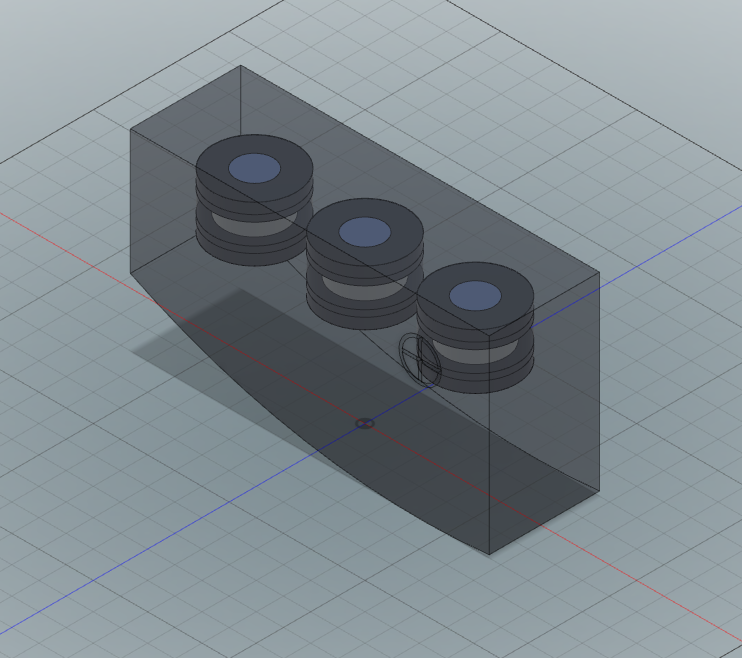
Figure 2: An illustration with translucency showing an SLN Profile CT Lung Nodule Phantom.
Assessment process:
- Scan the SLN Profile accepted Quantitative CT reference object (example in Fig. 2)
- Calculate metrics at two locations closest to iso-center and at 160.0 mm
- Use linear interpolation for 160.0 mm measurements
- Follow specific calculation methods for each metric
Note: Alternative protocols may be needed for small Field of View (FOV) scans.
Additional notes: A Modulation Transfer Function at a 50% cutoff frequency (MTF 50) value can be translated to an In-plane Point Spread Function sigma using the following equation [46]:
σxy = √(−2 ln m0) / (2πμ0)
where m0 is the MTF value and μ0 is the frequency. Thus, a conversion from gaussian PSF to MTF is:
m0 = e−(2πμ0σxy)2 / 2
More specifically, the conversion from PSF to MTF50 is:
m0 = e−(πσxy)2 / 2
5.1.2 Nodule Analysis Software Characteristics
Two metrics assess measurement quality:
- Measurement Bias: Deviation of mean value from true value for volumetric measurements
- Coefficient of Variation (CV): Ratio of standard deviation to mean for repeated measurements
Assessment methods:
- Measure volumes of geometric objects with known volumes
- Measure volumes of short-time interval repeat scans of nodules with varying characteristics
References
- Henschke CI, Yankelevitz DF, Libby DM, Pasmantier MW, Smith JP, Miettinen OS. Survival of patients with stage I lung cancer detected on CT screening. N Engl J Med. 2006; 355(17):1763-71.
- Aberle DR, Adams AM, Berg CD, et al. Reduced lung-cancer mortality with low-dose computed tomographic screening. N Engl J Med. 2011; 365(5):395-409.
- Yankelevitz DF, Reeves AP, Kostis WJ, Zhao B, Henschke CI. Small pulmonary nodules: volumetrically determined growth rates based on CT evaluation. Radiology. 2000; 217(1):251-6.
- Bolte H, Jahnke T, Schafer FK, et al. Interobserver-variability of lung nodule volumetry considering different segmentation algorithms and observer training levels. Eur J Radiol. 2007; 64(2):285-95.
- Gierada DS, Pilgram TK, Ford M, et al. Lung cancer: interobserver agreement on interpretation of pulmonary findings at low-dose CT screening. Radiology. 2008; 246(1):265-72.
- van Klaveren RJ, Oudkerk M, Prokop M, et al. Management of lung nodules detected by volume CT scanning. N Engl J Med. 2009; 361(23):2221-9.
- Singh S, Pinsky P, Fineberg NS, et al. Evaluation of reader variability in the interpretation of follow-up CT scans at lung cancer screening. Radiology. 2011; 259(1):263-70.
- Petrick N, Kim HJ, Clunie D, et al. Comparison of 1D, 2D, and 3D nodule sizing methods by radiologists for spherical and complex nodules on thoracic CT phantom images. Acad Radiol. 2014; 21(1):30-40.
- Mulshine JL, Gierada DS, Armato SG, 3rd, et al. Role of the Quantitative Imaging Biomarker Alliance in optimizing CT for the evaluation of lung cancer screen-detected nodules. J Am Coll Radiol. 2015; 12(4):390-5.
- Das M, Muhlenbruch G, Katoh M, et al. Automated volumetry of solid pulmonary nodules in a phantom: accuracy across different CT scanner technologies. Invest Radiol. 2007; 42(5):297-302.
- Ravenel JG, Leue WM, Nietert PJ, Miller JV, Taylor KK, Silvestri GA. Pulmonary nodule volume: effects of reconstruction parameters on automated measurements--a phantom study. Radiology. 2008; 247(2):400-8.
- Goo JM, Tongdee T, Tongdee R, Yeo K, Hildebolt CF, Bae KT. Volumetric measurement of synthetic lung nodules with multi-detector row CT: effect of various image reconstruction parameters and segmentation thresholds on measurement accuracy. Radiology. 2005; 235(3):850-6.
- Chen B, Barnhart H, Richard S, Colsher J, Amurao M, Samei E. Quantitative CT: technique dependence of volume estimation on pulmonary nodules. Phys Med Biol. 2012; 57(5):1335-48.
- Larici AR, Storto ML, Torge M, et al. Automated volumetry of pulmonary nodules on multidetector CT: influence of slice thickness, reconstruction algorithm and tube current. Preliminary results. Radiol Med. 2008; 113(1):29-42.
- Xie X, Willemink MJ, de Jong PA, et al. Small irregular pulmonary nodules in low-dose CT: observer detection sensitivity and volumetry accuracy. AJR Am J Roentgenol. 2014; 202(3):W202-9.
- Willemink MJ, Leiner T, Budde RP, et al. Systematic error in lung nodule volumetry: effect of iterative reconstruction versus filtered back projection at different CT parameters. AJR Am J Roentgenol. 2012; 199(6):1241-6.
- Wielputz MO, Lederlin M, Wroblewski J, et al. CT volumetry of artificial pulmonary nodules using an ex vivo lung phantom: influence of exposure parameters and iterative reconstruction on reproducibility. Eur J Radiol. 2013; 82(9):1577-83.
- Chen B, Barnhart H, Richard S, Robins M, Colsher J, Samei E. Volumetric quantification of lung nodules in CT with iterative reconstruction (ASiR and MBIR). Med Phys. 2013; 40(11):111902.
- Wormanns D, Kohl G, Klotz E, et al. Volumetric measurements of pulmonary nodules at multi-row detector CT: in vivo reproducibility. Eur Radiol. 2004; 14(1):86-92.
- Goodman LR, Gulsun M, Washington L, Nagy PG, Piacsek KL. Inherent variability of CT lung nodule measurements in vivo using semiautomated volumetric measurements. AJR Am J Roentgenol. 2006; 186(4):989-94.
- Gietema HA, Schaefer-Prokop CM, Mali WP, Groenewegen G, Prokop M. Pulmonary nodules: Interscan variability of semiautomated volume measurements with multisection CT-- influence of inspiration level, nodule size, and segmentation performance. Radiology. 2007; 245(3):888-94.
- Rampinelli C, De Fiori E, Raimondi S, Veronesi G, Bellomi M. In vivo repeatability of automated volume calculations of small pulmonary nodules with CT. AJR Am J Roentgenol. 2009; 192(6):1657-61.
- de Hoop B, Gietema H, van Ginneken B, Zanen P, Groenewegen G, Prokop M. A comparison of six software packages for evaluation of solid lung nodules using semi-automated volumetry: what is the minimum increase in size to detect growth in repeated CT examinations. Eur Radiol. 2009; 19(4):800-8.
- Marchiano A, Calabro E, Civelli E, et al. Pulmonary nodules: volume repeatability at multidetector CT lung cancer screening. Radiology. 2009; 251(3):919-25.
- Ko JP, Berman EJ, Kaur M, et al. Pulmonary Nodules: growth rate assessment in patients by using serial CT and three-dimensional volumetry. Radiology. 2012; 262(2):662-71.
- Henschke CI, Yankelevitz DF, Yip R, et al. Tumor volume measurement error using computed tomography imaging in a phase II clinical trial in lung cancer. J Med Imaging (Bellingham). 2016; 3(3):035505.
- Das M, Ley-Zaporozhan J, Gietema HA, et al. Accuracy of automated volumetry of pulmonary nodules across different multislice CT scanners. Eur Radiol. 2007; 17(8):1979-84.
- Coenen A, Honda O, van der Jagt EJ, Tomiyama N. Computer-assisted solid lung nodule 3D volumetry on CT: influence of scan mode and iterative reconstruction: a CT phantom study. Jpn J Radiol. 2013; 31(10):677-84.
- ACR-STR. ACR-STR practice parameter for the performance and reporting of lung cancer screening thoracic computed tomography (CT). 2014.
- Honda O, Johkoh T, Sumikawa H, et al. Pulmonary nodules: 3D volumetric measurement with multidetector CT--effect of intravenous contrast medium. Radiology. 2007; 245(3):881-7.
- Rampinelli C, Raimondi S, Padrenostro M, et al. Pulmonary nodules: Contrast-enhanced volumetric variation at different CT scan delays. AJR Am J Roentgenol. 2010; 195(1):149-54.
- Toth T, Ge Z, Daly MP. The influence of patient centering on CT dose and image noise. Med Phys. 2007; 34(7):3093-101.
- Kaasalainen T, Palmu K, Reijonen V, Kortesniemi M. Effect of patient centering on patient dose and image noise in chest CT. AJR Am J Roentgenol. 2014; 203(1):123-30.
- Goo JM, Kim KG, Gierada DS, Castro M, Bae KT. Volumetric measurements of lung nodules with multi-detector row CT: effect of changes in lung volume. Korean J Radiol. 2006; 7(4):243-8.
- Petkovska I, Brown MS, Goldin JG, et al. The effect of lung volume on nodule size on CT. Acad Radiol. 2007; 14(4):476-85.
- Nietert PJ, Ravenel JG, Leue WM, et al. Imprecision in automated volume measurements of pulmonary nodules and its effect on the level of uncertainty in volume doubling time estimation. Chest. 2009; 135(6):1580-7.
- Gavrielides MA, Zeng R, Myers KJ, Sahiner B, Petrick N. Benefit of overlapping reconstruction for improving the quantitative assessment of CT lung nodule volume. Acad Radiol. 2013; 20(2):173-80.
- Willemink MJ, de Jong PA, Leiner T, et al. Iterative reconstruction techniques for computed tomography Part 1: technical principles. Eur Radiol. 2013; 23(6):1623-31.
- Willemink MJ, Borstlap J, Takx RA, et al. The effects of computed tomography with iterative reconstruction on solid pulmonary nodule volume quantification. PLoS One. 2013; 8(2):e58053.
- Revel MP, Lefort C, Bissery A, et al. Pulmonary nodules: preliminary experience with three-dimensional evaluation. Radiology. 2004; 231(2):459-66.
- Petrou M, Quint LE, Nan B, Baker LH. Pulmonary nodule volumetric measurement variability as a function of CT slice thickness and nodule morphology. AJR Am J Roentgenol. 2007; 188(2):306-12.
- Wang Y, van Klaveren RJ, van der Zaag-Loonen HJ, et al. Effect of nodule characteristics on variability of semiautomated volume measurements in pulmonary nodules detected in a lung cancer screening program. Radiology. 2008; 248(2):625-31.
- Hein PA, Romano VC, Rogalla P, et al. Linear and volume measurements of pulmonary nodules at different CT dose levels - intrascan and interscan analysis. Rofo. 2009; 181(1):24-31.
- Hein PA, Romano VC, Rogalla P, et al. Variability of semiautomated lung nodule volumetry on ultralow-dose CT: comparison with nodule volumetry on standard-dose CT. J Digit Imaging. 2010; 23(1):8-17.
- Gietema HA, Wang Y, Xu D, et al. Pulmonary nodules detected at lung cancer screening: interobserver variability of semiautomated volume measurements. Radiology. 2006; 241(1):251-7.
- Wang GE, Skinner MW, Vannier MW. Temporal Bone Volumetric Image Deblurring in Spiral Computed Tomography Scanning. Academic Radiology. 1995; 2:888-895.
Additional References
- Gavrielides MA, Li Q, Zeng R, Myers KJ, Sahiner B, Petrick N. Minimum detectable change in lung nodule volume in a phantom CT study. Acad Radiol. 2013; 20(11):1364-70.
- Bolte H, Riedel C, Jahnke T, et al. Reproducibility of computer-aided volumetry of artificial small pulmonary nodules in ex vivo porcine lungs. Invest Radiol. 2006; 41(1):28-35.
- Bolte H, Riedel C, Muller-Hulsbeck S, et al. Precision of computer-aided volumetry of artificial small solid pulmonary nodules in ex vivo porcine lungs. Br J Radiol. 2007; 80(954):414-21.
- Wang Y, de Bock GH, van Klaveren RJ, et al. Volumetric measurement of pulmonary nodules at low-dose chest CT: effect of reconstruction setting on measurement variability. Eur Radiol. 2010; 20(5):1180-7.
- Bolte H, Riedel C, Knoss N, et al. Computed tomography-based lung nodule volumetry--do optimized reconstructions of routine protocols achieve similar accuracy, reproducibility and interobserver variability to that of special volumetry protocols? Rofo. 2007; 179(3):276-81.
- de Jong PA, Leiner T, Lammers JW, Gietema HA. Can low-dose unenhanced chest CT be used for follow-up of lung nodules? AJR Am J Roentgenol. 2012; 199(4):777-80.
- Christe A, Torrente JC, Lin M, et al. CT screening and follow-up of lung nodules: effects of tube current-time setting and nodule size and density on detectability and of tube current-time setting on apparent size. AJR Am J Roentgenol. 2011; 197(3):623-30.
- Honda O, Sumikawa H, Johkoh T, et al. Computer-assisted lung nodule volumetry from multi-detector row CT: influence of image reconstruction parameters. Eur J Radiol. 2007; 62(1):106-13.
- Young S, Kim HJ, Ko MM, Ko WW, Flores C, McNitt-Gray MF. Variability in CT lung-nodule volumetry: Effects of dose reduction and reconstruction methods. Med Phys. 2015; 42(5):2679-89.
- Ashraf H, de Hoop B, Shaker SB, et al. Lung nodule volumetry: segmentation algorithms within the same software package cannot be used interchangeably. Eur Radiol. 2010; 20(8):1878-85.
- Christe A, Bronnimann A, Vock P. Volumetric analysis of lung nodules in computed tomography (CT): comparison of two different segmentation algorithm softwares and two different reconstruction filters on automated volume calculation. Acta Radiol. 2014; 55(1):54-61.
- Zhao YR, Ooijen PM, Dorrius MD, et al. Comparison of three software systems for semi-automatic volumetry of pulmonary nodules on baseline and follow-up CT examinations. Acta Radiol. 2013; 55(6):691-8.
- Gavrielides MA, Kinnard LM, Myers KJ, Petrick N. Noncalcified lung nodules: volumetric assessment with thoracic CT. Radiology. 2009; 251(1):26-37.
- Marten K, Engelke C. Computer-aided detection and automated CT volumetry of pulmonary nodules. Eur Radiol. 2007; 17(4):888-901.
- Boll DT, Gilkeson RC, Fleiter TR, Blackham KA, Duerk JL, Lewin JS. Volumetric assessment of pulmonary nodules with ECG-gated MDCT. AJR Am J Roentgenol. 2004; 183(5):1217-23.
- Snoeckx A, Silva M, Prosch H et al. Lung cancer screening with low-dose CT: definition of positive, indeterminate, and negative screen results. A nodule management recommendation from the European Society of Thoracic Imaging. Eur Radiol. 2025; 1-13.
Metrology References
- Obuchowski NA, Buckler A, Kinahan PE, Chen-Mayer H, Petrick N, Barboriak DP, Bullen J, Barnhart H, Sullivan DC. Statistical Issues in Testing Conformance with the Quantitative Imaging Biomarker Alliance (ALA/QIBA) Profile Claims. Acad Radiol. 2016; 23(4):496-506.
- Kessler LG, Barnhart HX, Buckler AJ, et al. The emerging science of quantitative imaging biomarkers: terminology and definitions for scientific studies and for regulatory submissions. SMMR 2015; 24(1): 9-26.
- Raunig D, McShane LM, Pennello G, et al. Quantitative imaging biomarkers: a review of statistical methods for technical performance assessment. SMMR 2015; 24(1): 27-67.
- Obuchowski NA, Reeves AP, Huang EP, et al. Quantitative Imaging Biomarkers: A Review of Statistical Methods for Computer Algorithm Comparisons. SMMR 2015; 24(1): 68-106.
Appendices
Appendix A: Discussion on Clinical Context
- Low-dose CT benefits:
- Nodule measurement:
- Challenges in lung cancer CT screening:
- Balancing accurate nodule detection and characterization with radiation exposure risks
- Optimizing protocols for long-term screening (potentially over two decades)
- Profile aims:
- Establish confidence levels for measuring nodule volume and changes
- Quantifies nodule volume and volume doubling time to support standardized nodule management per European Society of Thoracic Imaging recommendations [62]
- Provide specifications for users and equipment developers
- Target various stakeholders involved in lung cancer screening
- Intended audience:
- Healthcare professionals (radiologists, technologists, physicists)
- Equipment manufacturers and software developers
- Biopharmaceutical companies and clinical trialists
- Policy makers and administrators
- Relevance:
- Primarily for asymptomatic persons in CT screening programs
May also apply to patients with known or incidentally-detected solid pulmonary nodules (6–10 mm diameter range).
Appendix B: Clinical Interpretation Examples
Claim 1: Nodule Volume
The true size of a nodule is defined by the measured volume and the 95% confidence intervals. These intervals represent the range within which the true volume likely falls.
Example 1:
- Measured volume: 150 mm3 (6.6 mm diameter)
- 95% confidence interval: 65 mm3 to 235 mm3 (5.0 mm to 7.7 mm diameter)
Example 2:
- Measured volume: 500 mm3 (9.8 mm diameter)
- 95% confidence interval: 343 mm3 to 657 mm3 (8.7 mm to 10.8 mm diameter)
Example 3:
- Measured volume: 800 mm3 (11.5 mm diameter)
- 95% confidence interval: 612 mm3 to 988 mm3 (10.5 mm to 12.4 mm diameter)
Claim 2: Nodule Volume Change
This claim helps determine if a change in nodule size is real or due to measurement variability.
Example 1:
- Baseline: 524 mm3 (10.0 mm diameter)
- Follow-up: 917 mm3 (12.0 mm diameter)
- Measured change: +75%
- Interpretation: Since 75% > 39%, we are 95% confident this is a real change.
- 95% confidence interval for true change: 149 mm3 to 637 mm3 increase
Example 2:
- Baseline: 180 mm3 (7.0 mm diameter)
- Follow-up: 270 mm3 (8.0 mm diameter)
- Measured change: +50%
- Interpretation: Since 50% < 80%, we cannot be confident this is a real change.
Note: These claims have been informed by clinical trial data, theoretical analysis, simulations, review of the literature, and expert consensus. They assume compliance with the Profile specifications and may be subject to refinement based on clinical implementation data. The claims have not yet been fully substantiated by studies that strictly conform to the specifications given here. It is expected that during implementation in the clinical setting, data on the actual performance will be collected and any appropriate changes made to the claims or the details of the Profile.
Appendix C: Acknowledgements and Attributions
This document is proffered by the Small Lung Nodule (SLN) Biomarker Committee. The group is composed of scientists representing academia, the imaging device manufacturers, image analysis tool software developers, image analysis laboratories, biopharmaceutical industry, government research organizations, professional societies, and regulatory agencies, among others. All work is classified as pre-competitive.
A description of the former QIBA SLN Biomarker Committee and its work can be found at the following web link: QIBA Small Lung Nodule Biomarker Committee Wiki page. Since the cessation of QIBA, the activities of the SLN Biomarker Committee moved to sponsorship of the American Lung Association (ALA). This is the largest lung disease advocacy organization in the United States with long standing and extensive support of lung imaging research both alone and in collaboration with the National Heart, Lung and Blood Institute of the National Institutes of Health. The SLN Biomarker Committee will continue its work on optimizing quantitative imaging processes for chest CT screening with the recently formed Quantitative Medical Imaging Coalition (QMIC), the European consortium, Strengthening the Screening of Lung Cancer in Europe (SOLACE) project and other relevant parties to inclusively make progress in the critical area.
Appendix D: CT Image Quality Conformance
See the separate Appendix D document.
Appendix E: Work In Progress (WIP) Information
See the separate Appendix E document.